Skip to content
RotaPlan

Plan precisely – before the first anchor is set.

RotaPlan™ is a planning system for rotator cuff repair: an interactive 3D model of the shoulder, its bone built automatically from the MRI, in which you classify the tear, place anchors and rehearse the repair before surgery.

  • Utility model registered (DPMA)
  • Made in Germany
  • Research prototype

This browser cannot show the 3D view.

0%
Footprint coverage0%

The animation loops. Drag the white handle or the slider to pull the tendon yourself.

Schematic illustration: the bone is from a public MRI dataset, the tendon is drawn. No patient data, no clinical limits.

Today, the plan lives in the surgeon’s head

Rotator cuff repair is one of the most common procedures in shoulder surgery. Yet it is still planned from a subjective reading of the MRI: surgeons scroll through 2D slices and assemble a 3D picture of the tear in their head.

Surgeons read the MRI as 2D slices…

…and assemble the 3D picture in their head.

From slices to a 3D model of the bone.

And a repair plan to explore before surgery.

Illustration on a public MRI dataset: the slices and the bone are the same shoulder. Not a patient plan.
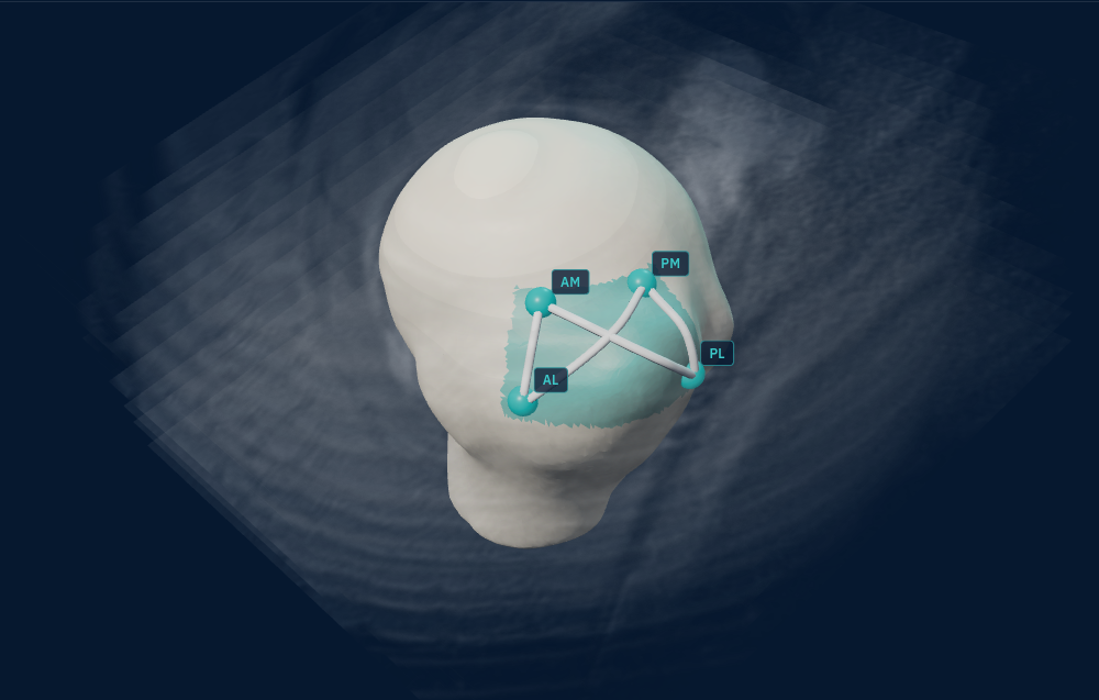
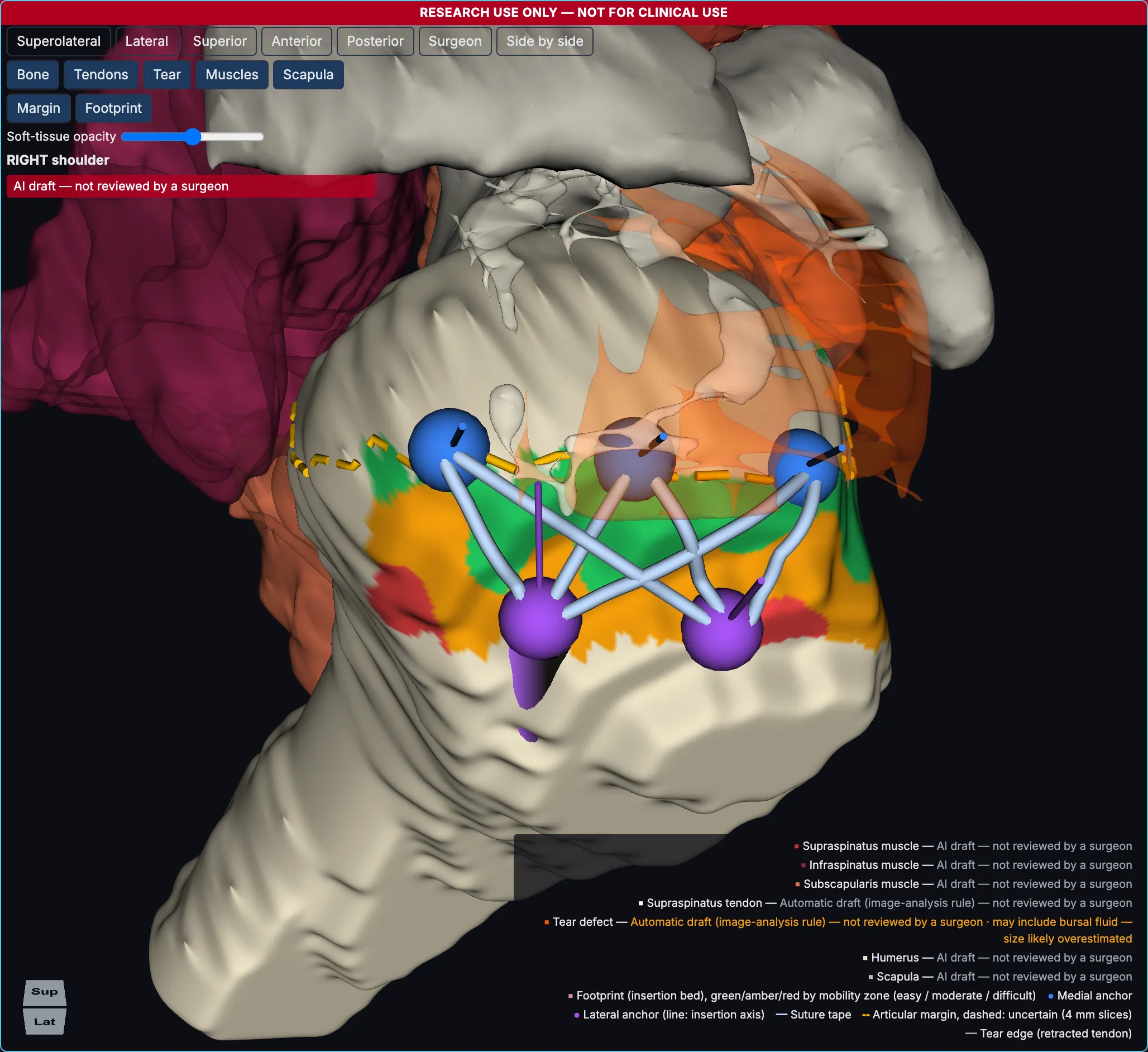
Illustration: the 3D humerus of a public MRI dataset among its own MRI slices, with an anchor plan (anchors AM, PM, AL, PL and crossing tapes) on the head
Illustration on a public MRI dataset: the slices and the bone are the same shoulder. Not a patient plan.
The real situation often only becomes clear during arthroscopy. And once the anchors are in, hindsight sets in: one more anchor, placed a little further apart, might have been the better choice.

Tear size, retraction and tissue quality all matter – but they are rarely measured in a reproducible way. To our knowledge, no commercial planning system exists for this soft-tissue reconstruction.

From MRI to repair plan in four steps

  1. Import MRI

    Load a de-identified shoulder MRI. RotaPlan checks image quality and finds the series each measurement needs.

  2. 3D model

    AI builds the bone model from the MRI and drafts the rotator cuff muscles, tendon and tear where the MRI allows. The surgeon reviews every draft.

  3. Tear analysis

    Size (Cofield) and retraction (Patte) are measured from the 3D model, fatty infiltration (Goutallier) where the muscles are segmented; pattern (Burkhart) and depth you set. You confirm or override every value.

  4. Explore & simulate

    Place anchors freely on the 3D humeral head and compare constructs – then send the signed plan to PACS.

You explore, you decide – RotaPlan makes the tear visible and testable in 3D.

What the prototype does today

3D shoulder modelRotate the humeral head freely and see tendon, tear edge and footprint in space – from anterior, lateral, superior or posterior.
RotaPlan 3D view of a humeral head with tendon, tear edge, footprint and a planned anchor construct, with muscles, tendon and tear as AI drafts not reviewed by a surgeon
Prototype screenshot on a de-identified research MRI. Muscles, tendon and tear are AI drafts, not reviewed by a surgeon.
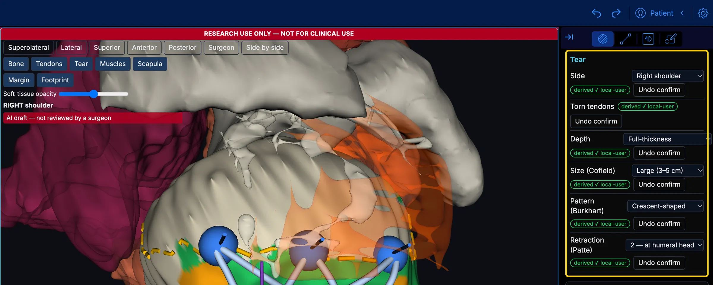
Standard grading, traceableCofield, Patte, Goutallier and Burkhart on one panel. Measured values come pre-filled and show where they came from; the surgeon confirms or overrides each one.
The RotaPlan app: 3D shoulder model beside the classification panel with pre-filled tear grading and the source of every value, with muscles, tendon and tear as AI drafts not reviewed by a surgeon
Prototype screenshot on a de-identified research MRI. Muscles, tendon and tear are AI drafts, not reviewed by a surgeon.
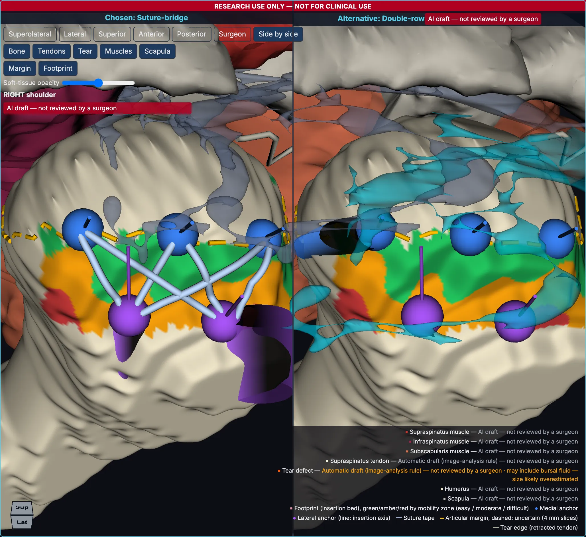
Compare constructsTwo anchor constructs side by side on the same shoulder – a double-row against a single-row repair, for example – each with its own warnings.
Two anchor constructs side by side on the same shoulder in RotaPlan, with muscles, tendon and tear as AI drafts not reviewed by a surgeon
Prototype screenshot on a de-identified research MRI. Muscles, tendon and tear are AI drafts, not reviewed by a surgeon.
Interactive simulatorPlace anchors and sutures, then watch the repair animation – also from the surgeon’s point of view.
Prototype screenshot on a de-identified research MRI. Muscles, tendon and tear are AI drafts, not reviewed by a surgeon.

Who it’s for

  • Shoulder surgeons

    Designed to support reproducible planning and a well-founded choice of technique – in 3D, before the first portal.

  • Hospitals & centres

    Standardised planning, a teaching mode for residents and the signed plan stored in PACS.

  • Medtech

    An implant-agnostic planning platform – open to partnership and licensing.

Where we are

RotaPlan is a working research prototype, protected by a utility model registered with the German Patent and Trade Mark Office, and not yet a CE-marked medical device. Next, we want to validate it on real cases – together with clinical partners.

  • Working prototype
  • Not a CE-marked medical device
  • Looking for clinical and industry partners

Founded by a shoulder surgeon and an ML engineer

  • Dr. Taha Yegani

    Dr. Taha Yegani

    Co-founder, medicine

    Specialist in orthopaedics with clinical experience in arthroscopic shoulder and knee surgery. Inventor of RotaPlan.

  • Dr. Mo Khatami

    Dr. Mo Khatami

    Co-founder, technology

    ML engineer, responsible for the AI pipeline and the software.

Partner with us

Are you a shoulder surgeon, a hospital or a medtech company and would like to evaluate or co-develop RotaPlan in research? Get in touch.